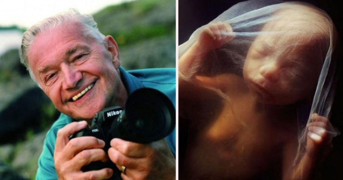
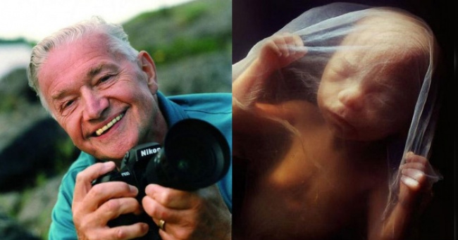
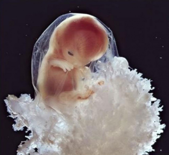
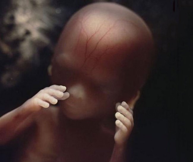
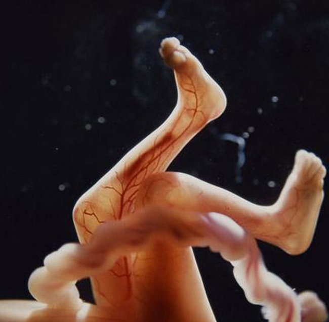
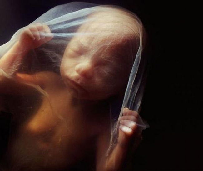
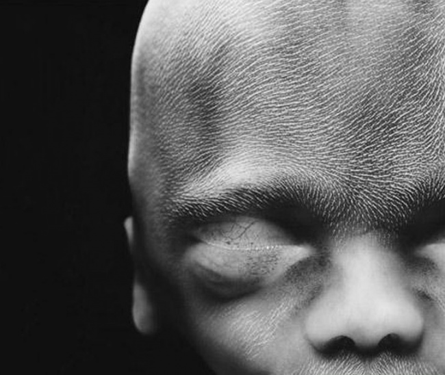
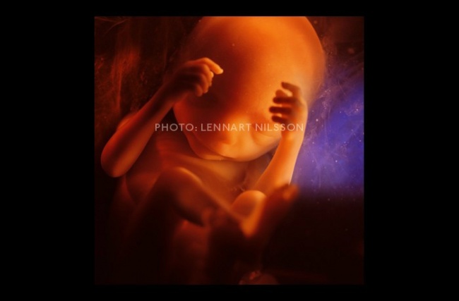
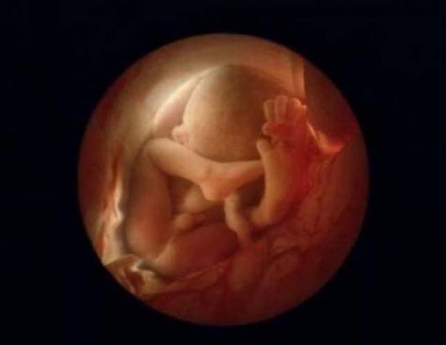
The world met first time to Lennart Nilsson in 1965, when his 16 pages photographs of human embryos published in LIFE magazine.
The shots were immediately reproduced in ’Stern’, ’Paris Match’, ’The Sunday Times’ and other publications. Nilsson was passionate towards Microscopes and the cameras since his childhood. His ambition was to show the world the beauty of the human body closely. He took his first photos of a fetus already in 1957, but they weren’t good enough to publish.
Nilsson succeeds to get his most accurate shots with the help of a cystoscope, a medical instrument which is used to examine the inside of the urinary bladder. He attached a tiny light with the camera to take the most perfect shots and took thousands of photos recording the life of the embryo in its mother’s womb.
LIFE magazine published Nilsson’s photographs with the title, The Drama of Life before Birth, a cover article of 16 pages containing Nilsson’s photographs. 8 million copies were sold out in a few days. The article is still among some of LIFE Magazine’s most important stories.
Nilsson produced something unbelievable, for the first time, people have seen the earliest development of human life with their own eyes.
Nilsson’s book, ’A Child is Born’, was published in 1965, and It sold out in just a few days and has been republished many times, it became one of the most popular photography books of all time.
Lennart Nilsson is now 91. He’s still fond of science and photography.
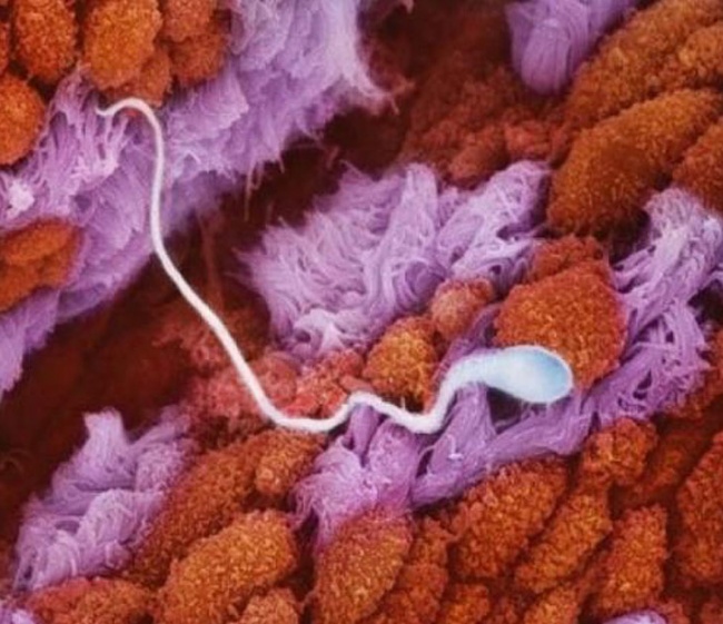
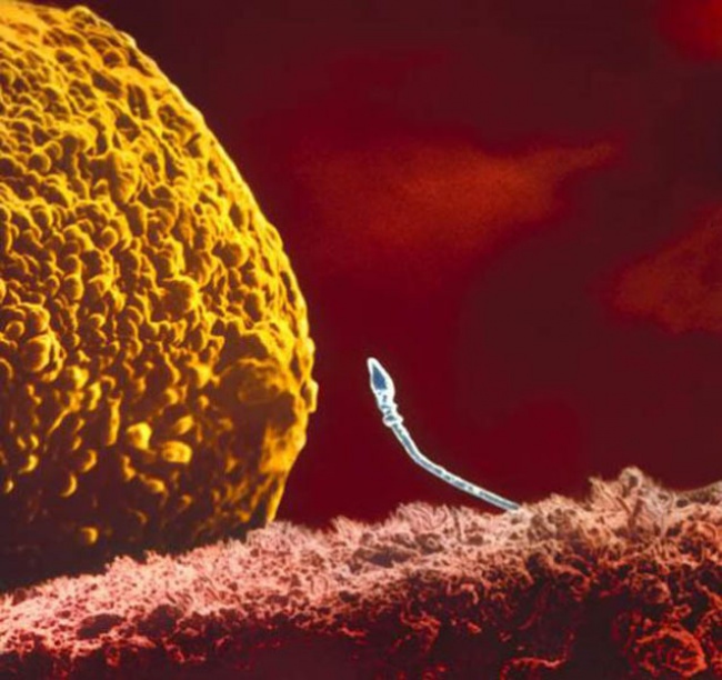
1 Sperm cell which has entered the fallopian tube
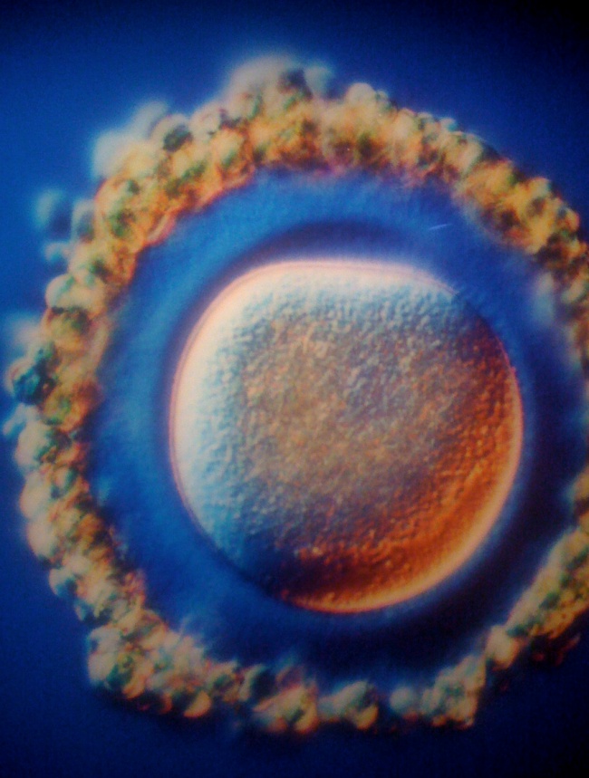
2 The egg
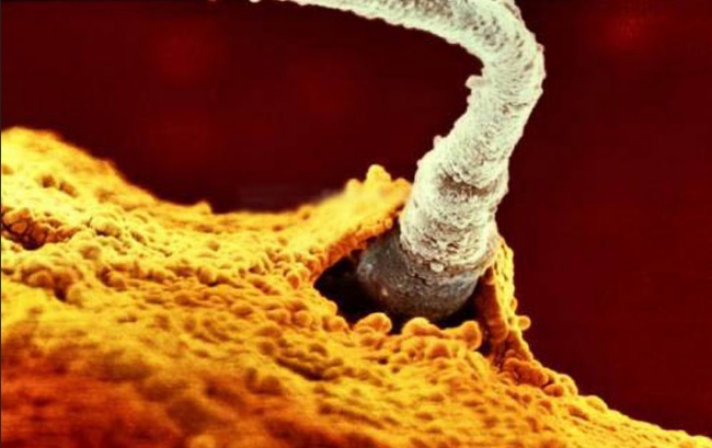
3 The crucial moment
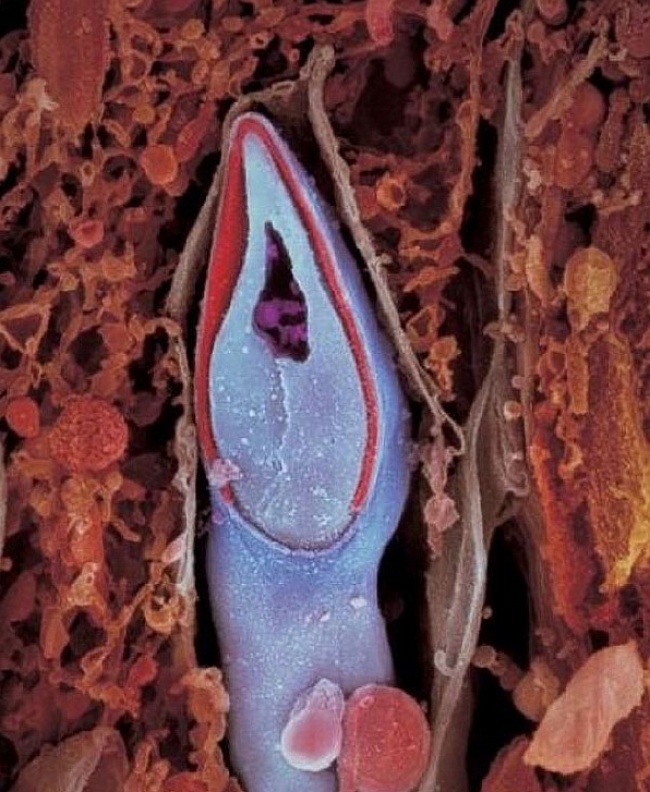
4 The winning sperm
5 The time when the egg cell and the sperm cell merged together
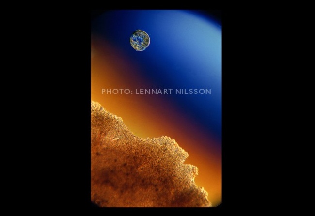
6 A week later, the embryo migrates to the womb by floating downwards through the fallopian tube
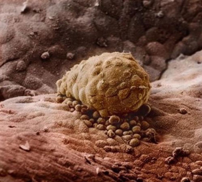
7 After 8 days. The zygote has attached itself to the wall of the uterus
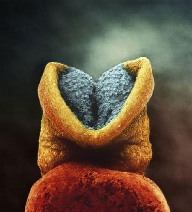
8 You can see the brain has started to develop
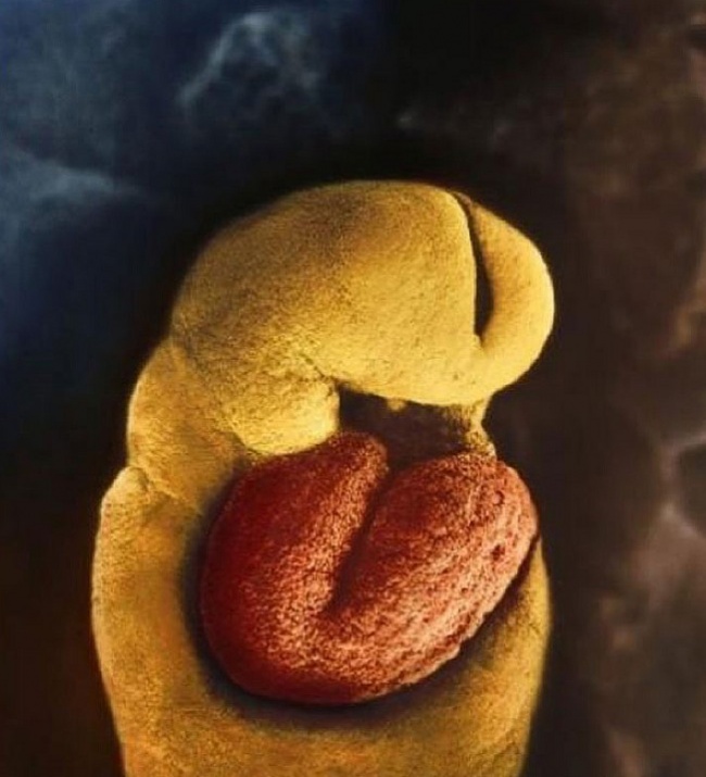
9 By the 18th day of development, the fetus’ heart begins to beat
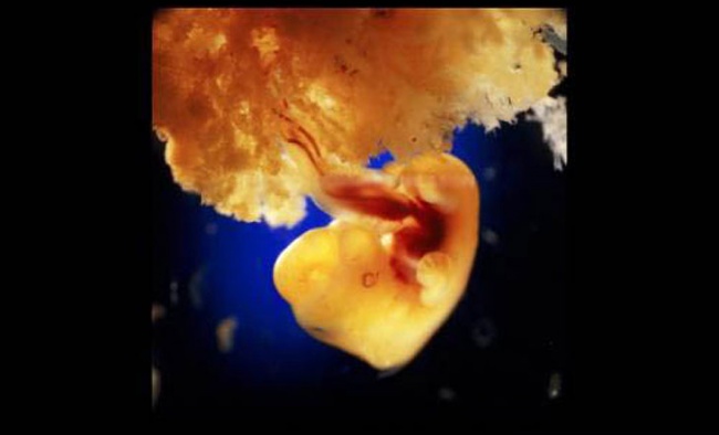
10 28 days after fertilization
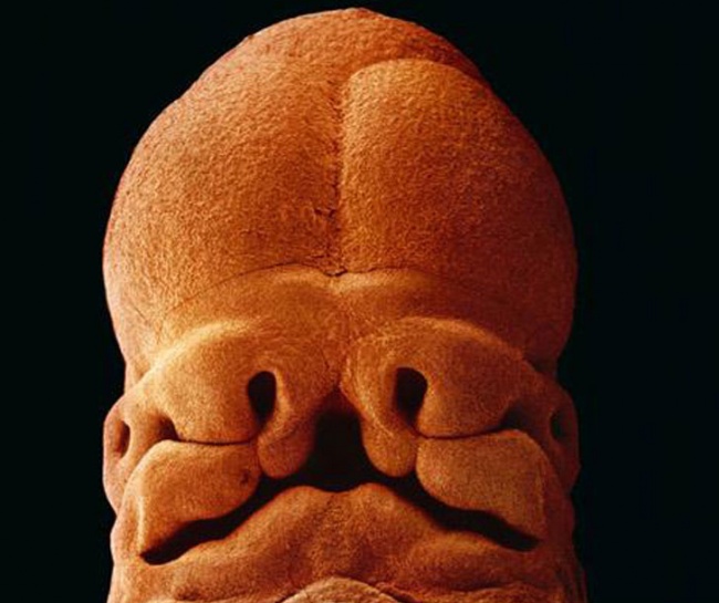
11 After 5 weeks. You can now distinguish the face with holes for eyes, nostrils and a mouth
12 40 days of development. The exterior cells of the fetus join with the loose surface of the uterus wall to form the placenta
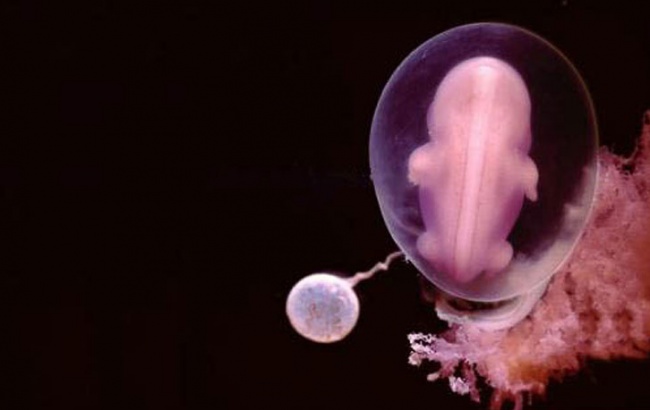
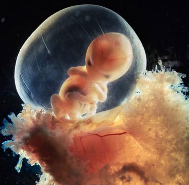
13 8-week-old embryo. The fetus is well protected in the fetal sac
14 10 weeks. Its eyelids are already half open
15 After 16 weeks
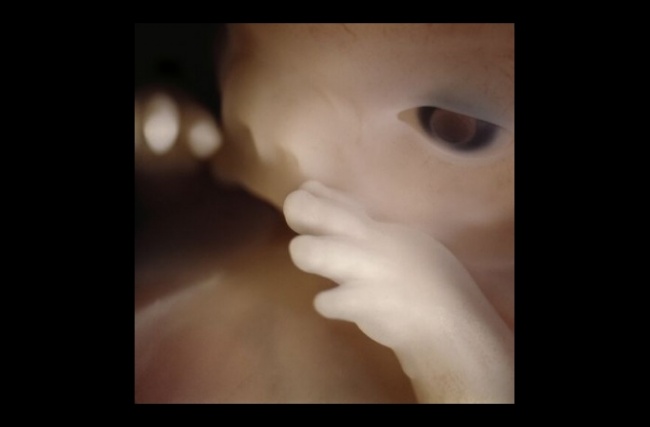
16 Fetus using its hands to explore its own body and surroundings
17 The skeleton, mainly consisting of flexible cartilage and a network of blood vessels, is visible through the skin
18 After 18 weeks
19 19 weeks
20 The fetus is now 20 centimeters long and hair starts growing
21 24 weeks
22 6 months
23 After 36 weeks, the child will see the world in a month


